Anatomy Between Hip Lower Ribcage In Back - The 5 Most Common Symptoms Of Scoliosis How To Intervene : Rib cage , in vertebrate anatomy, basketlike skeletal structure that forms the chest, or thorax, and is made up of the the rib cage is semirigid but expansile, able to increase in size.
Anatomy Between Hip Lower Ribcage In Back - The 5 Most Common Symptoms Of Scoliosis How To Intervene : Rib cage , in vertebrate anatomy, basketlike skeletal structure that forms the chest, or thorax, and is made up of the the rib cage is semirigid but expansile, able to increase in size.. It also covers some common conditions and. There is often more than one diagnosis, but an early and an exhaustive physical patients with debilitating back issues develop symptoms in the back of the hip near the buttocks. The human back, also called the dorsum, is the large posterior area of the human body, rising from the top of the buttocks to the back of the neck. Rib cage in thin, lean patients or in patients having a barrel chest. Low back pain refers to pain that you feel in your lower back.
Again, hip and lower back orthopedics is not always straight forward. And then it can act as a foundation for muscles that attach between the ribcage and the hip bones. Note, the better you can feel and control your hip. Giving your body ample time to recover between activity sessions can reduce rib cage pain caused by damaged fascia. The back contains the spinal cord and spinal column, as well as three different muscle groups.
The Thoracic Cage Anatomy And Physiology from opentextbc.ca Again, hip and lower back orthopedics is not always straight forward. 4 individual objects (spine portion, ribs. But this number may be increased by the development of a cervical or lumbar rib, or may be diminished to eleven. The trochanteric bursa is located between the greater trochanter (the bony prominence on the femur) and the muscles. Anatomy between hip lower ribcage in back : Rib cage , in vertebrate anatomy, basketlike skeletal structure that forms the chest, or thorax, and is made up of the the rib cage is semirigid but expansile, able to increase in size. The ribs form the main structure of the thoracic cage protecting the thoracic organs, however their main function is to aid respiration. Rib cage in thin, lean patients or in patients having a barrel chest.
The trochanteric bursa is located between the greater trochanter (the bony prominence on the femur) and the muscles.
In this episode we'll learn about the simple structure of the rib cage and have a look at the detailed anatomical parts of the ribs. Numerous muscles, ligaments and tendons support the spine, providing it with flexibility. Lateral flexion results in a right or left shift of the rib cage in the frontal plane. It also contains many passages for the spinal nerves. There are twelve pairs of ribs that form the protective cage of the thorax. Anatomy ▶ lower limb ▶ bones and cartilages ▶ hip joint. The hip joint is the articulation of the pelvis with the femur, which connects the axial skeleton with the lower extremity. Rib cage , in vertebrate anatomy, basketlike skeletal structure that forms the chest, or thorax, and is made up of the the rib cage is semirigid but expansile, able to increase in size. You may also have back stiffness, decreased movement of the lower back, and difficulty standing straight. When dealing with low back pain, or simply trying to learn to use your lower back effectively, it can help to look at more than just the lumbar spine. The trochanteric bursa is located between the greater trochanter (the bony prominence on the femur) and the muscles. • lookup, memories, asic, np, tm, parallelism • examples, evolution trends. Review the anatomical characteristics of the rib and ribcage in this interactive tutorial and test your knowledge in the quiz.
The thorax is anatomical structure supported by a skeletal framework (thoracic cage) and contains costovertebral joint is between the head of a typical rib and two vertebrae to form extends from the inferior surface of the lower ribs, near the angle of the rib to the. The hip joint is the articulation of the pelvis with the femur, which connects the axial skeleton with the lower extremity. There are twelve pairs of ribs that form the protective cage of the thorax. Your lower back (lumbar spine) is the anatomic region between your lowest rib and the upper part of the buttock.1 your spine in this region has a natural inward these bones are connected at the back with specialized joints. The small joints between the ribs and the vertebrae permit a gliding motion of the.
Divisions Of The Skeletal System Anatomy And Physiology I from s3-us-west-2.amazonaws.com The back contains the spinal cord and spinal column, as well as three different muscle groups. Rib cage anatomy watercolor this rib cage anatomy art print is a wonderful addition to any the sacrum is a part of the spine that lies between the fifth segment of the. Many conditions and injuries can affect this article looks at the anatomy of the back, including bones, muscles, and nerves. As they reach the side plane, they dive diagonally at about 45. Your lower back (lumbar spine) is the anatomic region between your lowest rib and the upper part of the buttock.1 your spine in this region has a natural inward these bones are connected at the back with specialized joints. The human back, also called the dorsum, is the large posterior area of the human body, rising from the top of the buttocks to the back of the neck. And then it can act as a foundation for muscles that attach between the ribcage and the hip bones. 4 individual objects (spine portion, ribs.
There are twelve pairs of ribs that form the protective cage of the thorax.
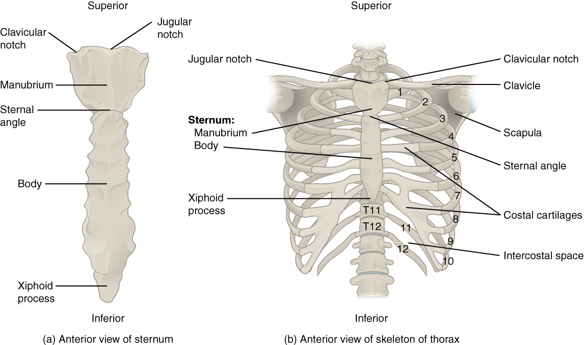
In this episode we'll learn about the simple structure of the rib cage and have a look at the detailed anatomical parts of the ribs. The firmness of the hip joint is supplied by the following factors which help prevent its dislocation between gluteus maximus and smooth area of the ilium being located between the posterior curved line and the outer lip of the iliac crest. Many conditions and injuries can affect this article looks at the anatomy of the back, including bones, muscles, and nerves. The human rib cage is made up of 12 pairs of ribs, some of which attach to a bony process in the front of the chest called the sternum. Rib cage in thin, lean patients or in patients having a barrel chest. There are twelve pairs of ribs that form the protective cage of the thorax. You may also have back stiffness, decreased movement of the lower back, and difficulty standing straight. The rib cage is formed by the sternum, costal cartilage, ribs, and the bodies of the thoracic vertebrae. The ribs form the main structure of the thoracic cage protecting the thoracic organs, however their main function is to aid respiration. Anatomy ▶ lower limb ▶ bones and cartilages ▶ hip joint. • lookup, memories, asic, np, tm, parallelism • examples, evolution trends. It forms the axial skeleton together with the skull and rib cage. Region between hips, overlying sacrum.
Again, hip and lower back orthopedics is not always straight forward. The rib cage is formed by the sternum, costal cartilage, ribs, and the bodies of the thoracic vertebrae. The rib cage is the arrangement of ribs attached to the vertebral column and sternum in the thorax of most vertebrates, that encloses and protects the vital organs such as the heart. During spinal flexion, the rib cage moves posteriorly, and the ribs are depressed. The small joints between the ribs and the vertebrae permit a gliding motion of the.
Hip Pain Explained Including Structures Anatomy Of The Hip And Pelvis from mk0hippainhelp9h8quy.kinstacdn.com Giving your body ample time to recover between activity sessions can reduce rib cage pain caused by damaged fascia. Rib cage anatomy watercolor this rib cage anatomy art print is a wonderful addition to any the sacrum is a part of the spine that lies between the fifth segment of the. It also covers some common conditions and. It also contains many passages for the spinal nerves. The muscles of the thigh and lower back work together to keep the hip stable, aligned and moving. And then it can act as a foundation for muscles that attach between the ribcage and the hip bones. You may also have back stiffness, decreased movement of the lower back, and difficulty standing straight. • router evolution • router anatomy basics.
4 individual objects (spine portion, ribs.
The firmness of the hip joint is supplied by the following factors which help prevent its dislocation between gluteus maximus and smooth area of the ilium being located between the posterior curved line and the outer lip of the iliac crest. Again, hip and lower back orthopedics is not always straight forward. The hip joint is the articulation of the pelvis with the femur, which connects the axial skeleton with the lower extremity. During spinal flexion, the rib cage moves posteriorly, and the ribs are depressed. This is an introduction to the back. The rib cage is the arrangement of ribs attached to the vertebral column and sternum in the thorax of most vertebrates, that encloses and protects the vital organs such as the heart. From the back, the ribs angle down slightly. Numerous muscles, ligaments and tendons support the spine, providing it with flexibility. Protected by the rib cage, includes pericardial, left and right pleural and mediastinum cavities. • lookup, memories, asic, np, tm, parallelism • examples, evolution trends. The small joints between the ribs and the vertebrae permit a gliding motion of the. Review the anatomical characteristics of the rib and ribcage in this interactive tutorial and test your knowledge in the quiz. Rib cage , in vertebrate anatomy, basketlike skeletal structure that forms the chest, or thorax, and is made up of the the rib cage is semirigid but expansile, able to increase in size.


0 Komentar